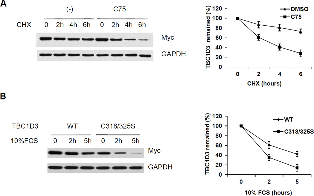
Figure 3. Palmitoylation influences TBC1D3 degradation.
(A) HeLa cells were transfected with Myc-TBC1D3. At 18 h after transfection, cells were treated with CHX (25µg/ml) with or without the fatty acid synthase inhibitor C75 (50µM). Cell extracts (10µg) were separated by SDS-PAGE followed by immunoblot analysis to monitor the level of TBC1D3. The graph shows the quantification from three experiments. (B) HeLa cells were transfected with wild-type and the palmitoylation-deficient TBC1D3 mutant -TBC1D3:C318/325S. The cells were starved for 4 h and stimulated with 10% FCS for the times indicated. Lysates were separated by SDS-PAGE. TBC1D3 levels were measured by immunoblotting with an anti-Myc antibody. The graph shows the quantification from three experiments.

