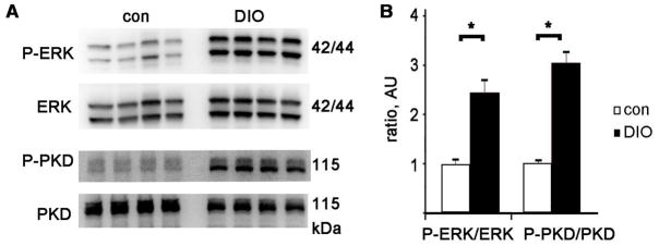
Figure 5. ERK and PKD are activated in DIO hearts.

A Representative immunoblots of phosphorylated ERK 1/2, the same membrane, stripped and reprobed with total ERK antibody, and below P-PKD (Ser916) and total PKD. B. Graph of P-ERK divided by total ERK and P-PKD divided by total PKD, normalized to control, arbitrary units. N= 8 each group, mean + SEM, * = p<0.01 by t-test
