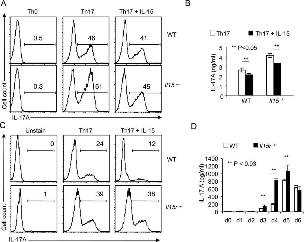
Figure 3. Genetic deficiency of IL-15 or IL-15 receptor increases IL-17A production in Th17 cultures.
(A) Flow cytometric histograms of IL-17A expression of naive WT (upper panel) or Il15−/− CD4 cells (lower panel) stimulated using anti-CD3 and anti-CD28 under non-polarizing conditions (Th0) or Th17 conditions for 96 hours with or without 20 ng/ml of IL-15. (B) ELISA quantification of IL-17A in supernatants from cells that were stimulated as in (A) under Th17 conditions (white bars) in the absence or presence of IL-15 (black bars). (C) Flow cytometric histograms of IL-17A expression of naive WT (upper panels) or Il15r−/− CD4 cells (lower panels) stimulated under Th0 or Th17 conditions as in (A). (D) ELISA quantification of IL-17A in supernatants from WT (white bars) or Il15r−/− (black bars) Th17 cells that were stimulated as in (C) at different days after stimulation.

