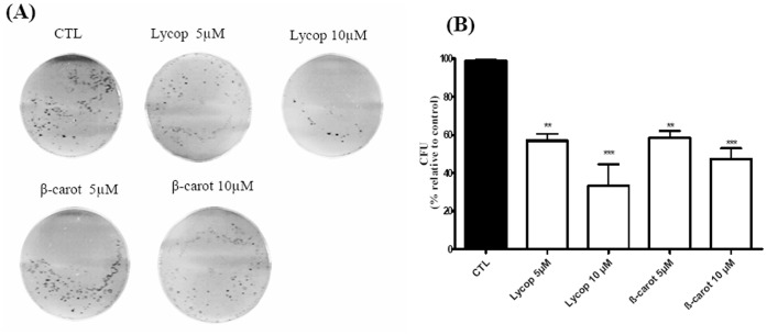
Figure 2. Formation of AtT-20 colonies.
The number of AtT-20 colonies was determined after 21 days of culture in DMEM supplemented with 10% FCS containing lycopene (Lycop) and beta-carotene (beta-carot) at concentrations of 5 and 10 µM. The number of colonies formed was detected by crystal violet staining. Phase contrast microscopy of AtT-20 cell colonies was observed on 6-well culture plates (A) and quantitative representation of the colonies formed (B). Data are presented as mean±standard deviation of 3 independent experiments, each performed at least in duplicate. *indicates significant differences from control group (**p<0.01, ***p<0.001).

