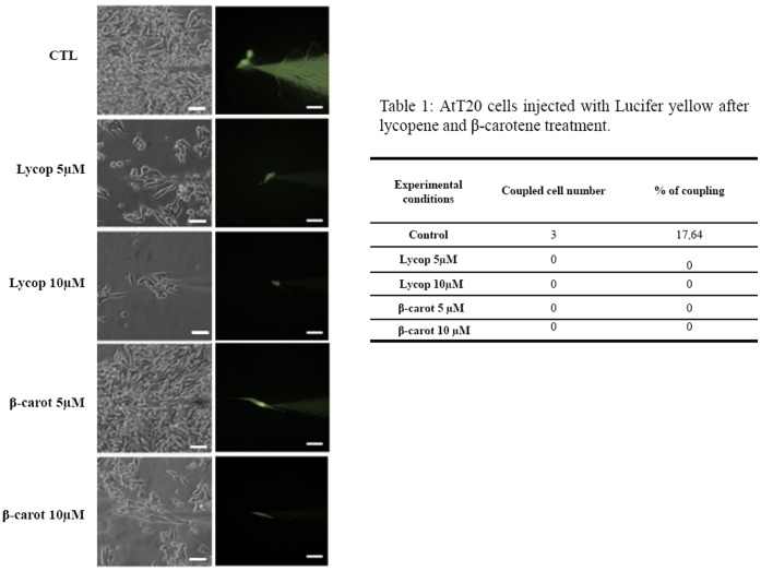
Figure 7. Photomicrographs from bright- and dark-field phase-contrast microscopy of AtT-20 cells treated with lycopene or beta-carotene 5 and 10 µM for 48 h and injected with Lucifer yellow iontophoretic dye (A).
A transfer of the dye occurred only in the control condition; the treatments completely blocked the dye transfer between neighboring cells. In B, the number of coupled cells and the rate of cell coupling (%). Phase-contrast images were taken from random fields. Bar: 80 µm.

