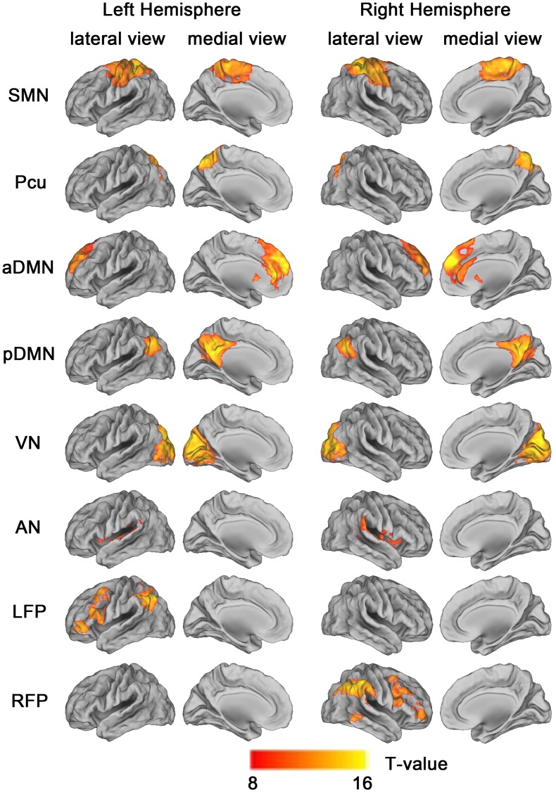
Figure 1. Cortical representation of the 8 resting state networks (RSNs) identified by independent component analysis.
Data are displayed on the lateral and medial surfaces of the left and right hemispheres of a brain surface map using CARET software [106]. The color scale represents T values in each RSN. Abbreviations: aDMN, anterior default mode network; AN, auditory network; LFP, left frontoparietal network; Pcu, precuneus network; pDMN, posterior default mode network; RFP, right frontoparietal network; SMN, sensorimotor network; VN, visual network.

