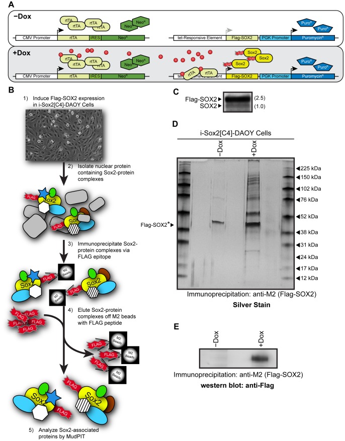
Figure 1. Engineering of iSOX2-DAOY medulloblastoma cells to identify SOX2-associated proteins.
(A) Schematic diagram of the lentiviral system used to introduce a Dox-inducible, epitope tagged SOX2 into DAOY MB cells. Two lentiviral vectors were used to introduce constitutively expressed reverse-tet transactivator (rtTA) and inducible (fs)SOX2 [labeled: Flag-SOX2]. (B) Protocol used to isolate SOX2-protein complexes from DAOY MB cells for downstream MudPIT analysis. (C) Western blot analysis probing for SOX2 with a Sox2 antibody. The level of (fs)SOX2 was compared to the level of endogenous SOX2, which was set to 1. (D) Silver stain demonstrating enrichment of proteins following the induction of (fs)SOX2 with Dox and immunoprecipitation with M2-beads. The prominent band observed at ∼45 kDa is a common contaminant when M2-beads are used for immunoprecipitation. *The estimated position of (fs)SOX2 is indicated by an arrowhead. (E) Western blot analysis probing for Flag-SOX2 to verify immunoprecipitation using M2-beads.

