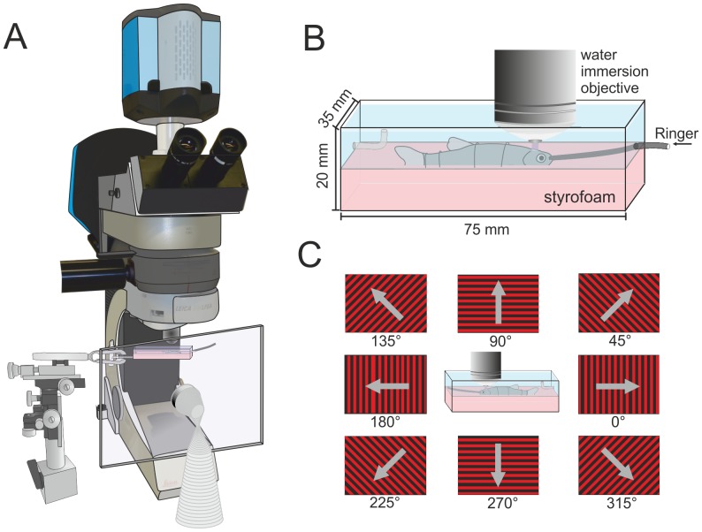
Figure 1. Schematic drawing of the experimental set-up.
A. The animal was placed under a standard upright fixed-stage microscope, equipped with a sensitive electron-multiplying CCD camera and LED-based epifluorescence illumination. To deliver visual stimuli, we used a TFT screen, which was placed in front of the eye facing the experimenter (drawn transparent for clarity). B. Detailed view of the recording chamber. Fish were placed on a styrofoam support tray and held in place by tungsten wires bent to support the animals. The chamber was continuously perfused with Hickmann ringer. C. Schematic display of the grating stimulus used for the determination of orientation and directional selectivity of tectal cells. For all data the denomination of the motion directions corresponds to the one shown here.

