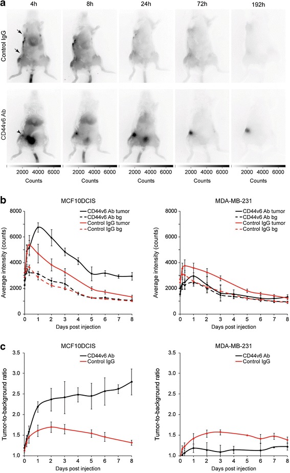
Fig. 1.

Noninvasive optical molecular imaging of breast cancers. a Representative SCID Beige mice bearing orthotopically transplanted MCF10DCIS (inguinal) and MDA-MB-231 (thoracic) tumors. Mice were intravenously injected in the tail vein with CD44v6 Ab or control IgG. At 4 h postinjection, tumor accumulation of CD44v6 Ab was observed in the MCF10DCIS tumors (arrowhead), whereas no accumulation of control IgG was observed in MCF10DCIS or MDA-MB-231 tumors (arrows). b Fluorescence intensity of MCF10DCIS tumors (left panel) or MDA-MB-231 tumors (right panel) and background of mice injected with CD44v6 Ab or control IgG over time. Data are displayed as average ± SEM (n = 6). (bg = background). c Tumor-to-background ratio of CD44v6 Ab and control IgG in MCF10DCIS and MDA-MB-231 tumors. Data are displayed as average ± SEM (n = 6).
