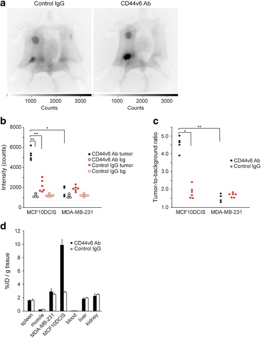
Fig. 2.

Intraoperative imaging of breast cancers. a Representative intraoperative fluorescence images of mice bearing MDA-MB-231 tumors and MCF10DCIS tumors 8 days postinjection with control IgG and CD44v6 Ab. Clear accumulation of CD44v6 Ab was observed in the MCF10DCIS tumor compared to control IgG. Higher signals in both tumors compared to the background were found, independent of the injected antibody due to enhanced perfusion and retention of the tumor. b Fluorescence intensity of MCF10DCIS and MDA-MB-231 tumors and the corresponding background (bg) in individual mice (*p < 0.05; **p < 0.01). c Tumor-to-background ratio of CD44v6 Ab and control IgG in MCF10DCIS and MDA-MB-231 tumors displayed for individual mice (*p < 0.05; **p < 0.01). d Biodistribution of CD44v6 Ab and control IgG 8 days postinjection. Tissue levels are expressed as percentage injected dose per gram tissue (%ID/g) as average ± SEM (n = 6).
