
Figure 1.
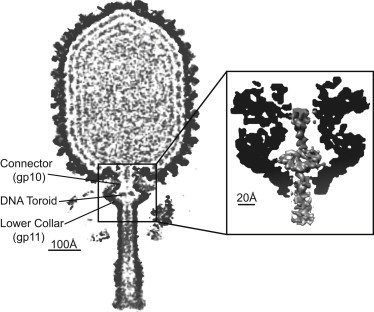
Cross section of the three-dimensional cryo-EM ϕ29 reconstruction (11). The virion map (purple, color online) is overlaid with the emptied map to highlight the genome. (Inset) Region to be simulated; the toroid constrained in the connector/lower collar cavity. Density maps generated with UCSF CHIMERA (36).