
Fig. 1.
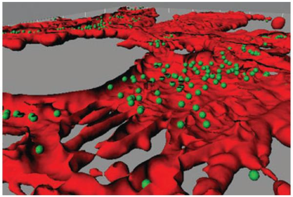
Invasion of human epithelial cells with Porphyromonas gingivalis. Confocal image of gingival epithelial cells [stained with TRITC-phalloidin (red)] infected with P. gingivalis [stained with FITC (green)]. The image was analyzed using Imaris version 5.0.1 software. A Z-stack of the x–y sections was converted to composite images using the iso surface and spot detection functions of the surpass option. The section view in the x and y axes was created using the clipping function. The image was generated by Masae Kuboniwa, Osaka University, Japan.