
Fig. 2.
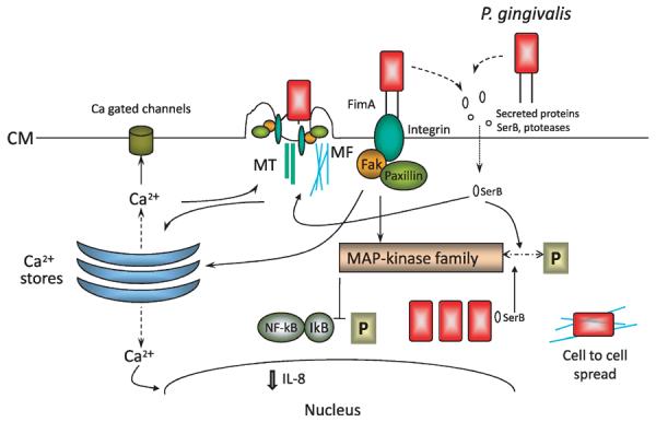
Model of interactions between Porphyromonas gingivalis and gingival epithelial cells that are associated with internalization. Proximity to gingival epithelial cells induces P. gingivalis to secrete proteins such as the SerB serine phosphatase. SerB enters gingival epithelial cells where it dephosphorylates target proteins, including mitogen-activated protein kinase family members, which in turn prevent NF-κB activation. SerB activity culminates in a reduction of interleukin-8 production and in the remodeling of microfilament and microtubule cytoskeletal architecture. Adhesion of P. gingivalis is mediated by the long (FimA) fimbriae that engage integrins and induce the formation of focal adhesin complexes and integrin-dependent signaling. Calcium ions (Ca2+) are released from intracellular stores, a signaling event that also funnels through the cytoskeletal structure, and the cytoskeletal re-arrangements allow P. gingivalis to enter the host cell. P. gingivalis cells rapidly locate in the perinuclear area where they replicate and utilize microfilaments to spread to adjacent gingival epithelial cells. CM, cytoplasmic membrane; IL-8, interleukin-8; IκB, inhibitor of κB; MAP, mitogen-activated protein kinase; MF, micro-filament; MT, microtubule; NF-κB, nuclear factor-κB; P, phosphate.