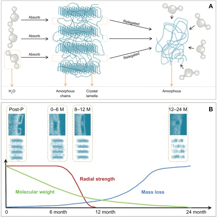
Figure 1.
Schematic representation of the poly-L-lactic acid bioresorption process. (A) Crystal lamella, interconnected by amorphous chains, absorbs water from the surrounding tissue after implantation in the coronary lesion. The amorphous polymer is more susceptible to hydration than the semicrystalline polymer. (B) Schematic representation of changes in the radial strength, molecular weight, and mass of the scaffold with time.
Note: The optical coherence tomography and corresponding molecular images, illustrate the changes in strut appearance and composition of the scaffold with time. Abbreviations: M, months; P, procedure.

