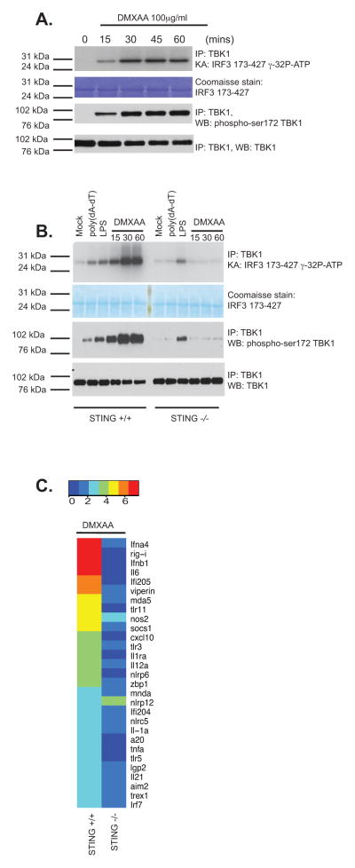
Figure 1. DMXAA signals via STING in macrophages.
(A) Wild-type immortalized bone marrow-derived macrophages were stimulated with DMXAA (100μg/ml) at the indicated time points. Whole cell lysates were prepared and endogenous TBK1 immunoprecipitated (IP) and analyzed by in vitro kinase assay and western blotted (WB) for phospho-TBK1 (Ser172) and total TBK1. (B) Wild-type or STING−/− bone marrow-derived macrophages were stimulated with poly(dA-dT) (3μg/ml), LPS [10ng/ml] or DMXAA (75μg/ml) as indicated and analyzed as in A. (C) BMDM from wild-type and STING−/− mice were stimulated with DMXAA (75μg/ml) for 4 hours and mRNA levels for a selection of innate immune genes were analyzed by Nanostring analysis. Heatmaps representing differentially regulated genes are presented and scaled by log2(X-min(X) + 1). Data are presented from one experiment which is representative of three experiments (A, B) or two experiments (C).

