
Figure 2.
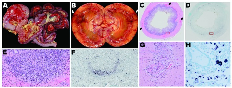
Organ tissues from sentinel dog (dog 1) and 2 other dogs (dogs 2 and 3) that were positive by in situ hybridization (ISH) analysis for dog circovirus (DogCV). A) Gross view of the gastrointestinal system from dog 1. Multifocal to coalescing hemorrhages are shown in the stomach and intestinal serosa. B) Gross view of the kidney from dog 2. Segmental regions of hemorrhage and necrosis (infarction) can be seen within the cortex and radiating from the medullary papilla to the capsule. C) Hemolyxin and eosin (H&E) stain of the ileum from dog 1. Peyer’s patches are moderately depleted, and multifocal regions of hemorrhage are present within the muscular intestinal wall. D) ISH of the ileum from dog 1. Peyer’s patches circumferentially contain abundant DogCV DNA. E) H&E stain of a Peyer’s patch in the ileum from dog 1, showing moderate depletion of lymphocytes and an increased population of macrophages within the germinal center and the peripheral base of the follicle. F) ISH of a Peyer’s patch in the ileum from dog 1. DogCV DNA is rich within the cytoplasm of abundant cells at the periphery of the follicle, and individual cells are scattered within the germinal center, lymphatic channels, and submucosa. The positive cells have the morphologic appearance of macrophages. G) H&E stain of the jejunum from dog 3, showing segmental, circumferential, fibrinoid necrosis of the artery. H) ISH of a mesenteric lymph node from dog 2. Macrophages in the medullary sinus and lymphatic cords contain abundant DogCV DNA.