
Figure 3.
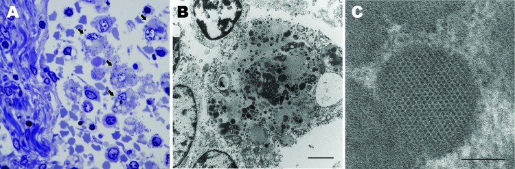
Lymph node from sentinel dog from which dog circovirus was identified. A) Toluidine blue stain shows multiple macrophages within the medullary sinus contain vacuoles and discrete, oblong to round, variably stained cytoplasmic bodies (arrows). B) A single macrophage adjacent to a lymphocyte (upper left) and partial profiles of other cells. Intracytoplasmic inclusion bodies are distributed throughout the macrophage cytoplasm, along with mitochondria and vacuoles. Scale bar indicates 2 µm. C) Intracytoplasmic inclusion bodies contain granular content and sometimes paracrystalline to herringbone arrays of 10–11 nm diameter viral-like particles. Scale bar indicates 100 nm.