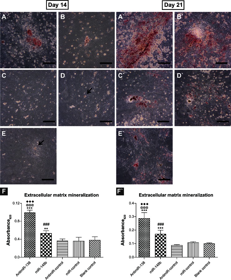
Figure 10.
Extracellular matrix mineralization mediated by anti-miR-138 and miR-148b. Transfected mesenchymal stem cells were stained using Alizarin red after 14 and 21 days of culture in osteogenic medium. (A and A`) Reverse transfection formulation of anti-miR-138, (B and B`) reverse transfection formulation of miR-148b, (C and C`) reverse transfection formulation of anti-miR-control, (D and D`) reverse transfection formulation of anti-miR-control and (E and E`) blank control tissue culture plate. The black arrows in (D and E) indicate tiny mineralized dots. Scale bar in micrographs represents 100 μm. (F and F`) Quantitative colorimetric results.
Notes: **P < 0.01 and ***P < 0.001 versus blank control tissue culture plate; ###P < 0.001 versus miR-control group; @@@P < 0.001 versus anti-miR-control group; ♦♦♦P < 0.001 versus miR-148b group.

