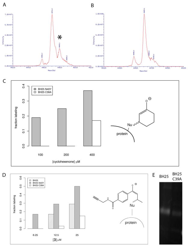
Figure 2.
Cyclohexenone and 3 labeling of BH25 and variants by HPLC/MS and fluorescent in-gel visualization. (A) Cyclohexenone labeling of BH25 N43Y measured by ESI MS and (B) labeling of the cysteine nucleophile knockout BH25 C39A. Both A and B were labeled at 200 μM cyclohexenone and 1 hour. Cyclohexenone at 200 μM labels specifically on the BH25 design (peak position marked by *), but not on the BH25 C39A knockout. (C) Specific labeling is also confirmed for 100 μM of cyclohexenone, but at a higher concentration of 400 μM a non-specific labeling is detectable in protein MS. (D) 3 labels BH25 N43Y specifically already at a very low concentration of 6.25 μM. At 12.5 μM the BH25 is labeled as well, however a small detectable peak arises also for the non-specific 3 labeling for the cysteine to alanine mutant (nucleophile knockout). (E) Specific binding of 3 is also confirmed for BH25 by in lysate “click” chemistry attachment of TAMRA dye (carboxytetramethyl-rhodamine) onto the free alkyne of the substrate. BH25 shows detectable labeling at 200 μM of 3 and 48 μM of TAMRA dye, while the cysteine to alanine mutant is labeled only weakly.

