
Figure 1. Arrested embryonic development in the absence of L3mbtl2.
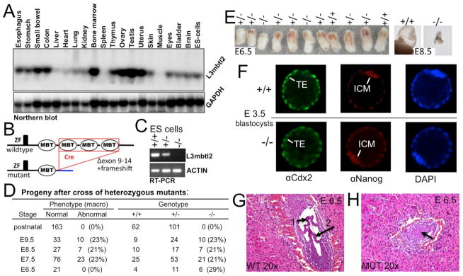
(A) L3mbtl2 is widely expressed. Northern blot analysis of RNA from selected tissues. (B) Illustration of L3mbtl2 conditional allele (for details see Suppl. Fig. S1). (C) RT-PCR analysis of mRNA for L3mbtl2 using primers in the 5-prime region (not deleted in the genome) demonstrates that mutant mRNA is non-detectable and likely unstable (see Fig. 2A for analysis of protein). (D, E) Progeny of heterozygous mutant mice bearing germline-excised L3mbtl2. (D) No homozygous mutants were detected after birth. At macroscopic examination upon dissection at E6.5, L3mbtl2−/− embryos appeared roughly normal in size (E, left). Following E7.5, mutants were growth retarded. At E8.5, mutant embryos were minute (E, right), and at E9.5, only debris was recovered from decidua. (F) Immunohistological analysis of L3mbtl2+/+ and L3mbtl2−/− blastocysts at E3.5 revealed that both form trophectoderm (TE, expressing Cdx2) and inner cell mass (ICM, expressing Nanog). (F, G) Histologic sections from uteri at E6.5 show wildtype embryos with well-defined cavities segmented by chorion (F, arrow 1) and amnion (F, arrow 2) membranes and mutant embryos with unstructured cores (G, arrow) (for further histology see Suppl. Fig. S2).