
Figure 6. Entry-side DNA movement occurs after 7 bp of DNA translocation towards the exit side and proceeds in 3-bp steps.
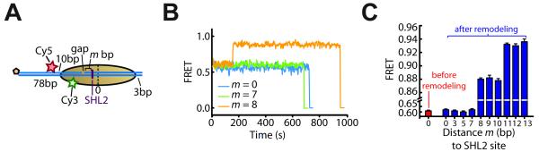
(A) Schematic of the nucleosome constructs used to monitor DNA movement at the entry side when exit-side translocation is restricted by a 2-nt ssDNA gap. The gap is located m bp away from the SHL2 site (shown as a purple line) such that m bp of DNA can be translocated to the exit side.
(B) FRET time traces of single m = 0, 7, and 8 bp nucleosomes (blue, green, and orange line, respectively) after addition of 12 nM ISW2 and 2 μM ATP at time zero.
(C) FRET values before (red bar) and after (blue bars) remodeling by ISW2 as a function of the distance m to the SHL2 site. Because the DNA path on the entry side may involve bending and/or twisting due to the direct interaction with the remodeling enzyme, we do not expect a similar linear dependence of FRET on the linker DNA length as on the exit-side where the linker DNA is largely free of enzyme-induced distortion. Data are shown as the mean ± SEM (N = 80 - 150 nucleosomes).
See also Figure S6.