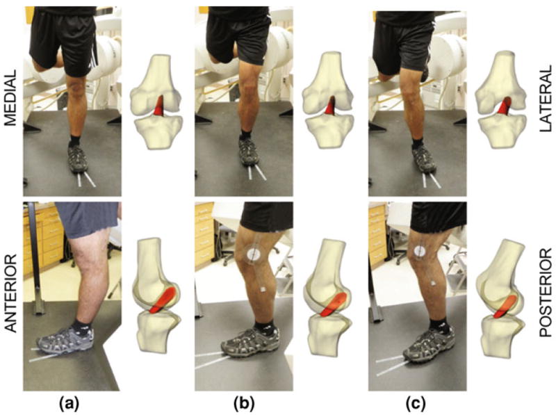
FIGURE 2.

Each subject was imaged using biplanar fluoroscopy. Using 3D MR-based models of the knee joint, we then reproduced the motion of each subject’s knee during testing as demonstrated in coronal (top) and sagittal views (bottom). These models were used to measure ACL length and joint kinematics in each of the three positions: (a) full extension, (b) 30° flexion, (c) a pose mimicking a valgus collapse position, with 30° flexion, 10° external rotation of tibia, and internal rotation of the hip.
