
Figure 2. see also Figure S2. A mouse model distinguishing between cystitis and pyelonephritis.
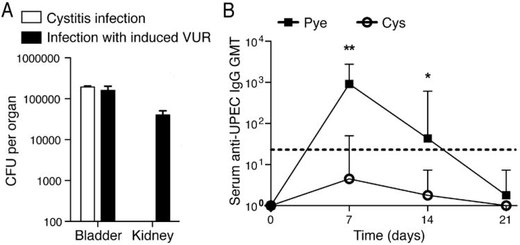
(A) Similar bacterial CFUs are obtained from bladders during both methods of infection; bacteria are only cultured from kidneys of pyelonephritis-infected mice. All data are representative of 2 independent experiments, n=3-4 per time point; error bars represent ±SEM. (B) Only mice with acute pyelonephritis show significant serum anti-UPEC IgG geometric mean titers (GMT) following infection. Dotted line denotes threshold of detection. *p<0.05; **p<0.01. Error bars represent the 95% confidence level; n=4-6.