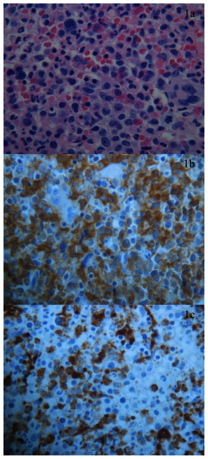
Figure 1.
BM biopsy, high magnification (x 40) shows prominent myeloid proliferation (a); elements with myelomonocytic morphology expressing CD33 (b) and CD68RPGM1 (c) are prevalent. Dysplastic granulocytopoiesis and megakaryocytopoiesis are evident. Cells with undifferentiated immature morphology or blast equivalent as monoblast or promonocytes are very rare.

