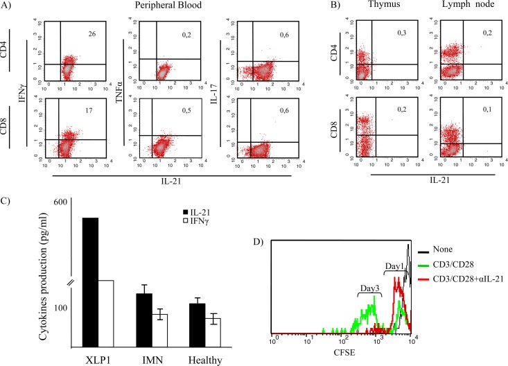
Fig 3.
Analysis of cytokine expression by T cell subtypes from the XLP-1 patient. IFN-γ was coexpressed by 26% of CD4+ T cells and 17% of CD8+ lymphocytes producing IL-21. (A) Peripheral blood T cells from the XLP-1 patient did not produce IL-17A. (B) Expression of IL-21 was not detected in single positive thymocytes or in lymph node T cells obtained from the XLP-1 patient in postmortem studies. (C) IL-21 and IFN-γ production levels by T cells from the XLP-1 patient, IMN patients (n = 7), and healthy individuals (n = 7) were measured in culture supernatants following stimulation with anti-CD3/CD28-coupled beads for 24 h. Results from the XLP-1 patient are expressed as the means of results from triplicate cultures; in the case of IMN patients and healthy individuals, results represent the means ± standard deviations (SD) obtained from 7 independent experiments. Neutralizing anti-IL-21 MAb inhibited T cell proliferation in all individuals tested. (D) Results obtained from the XLP-1 patient are shown.

