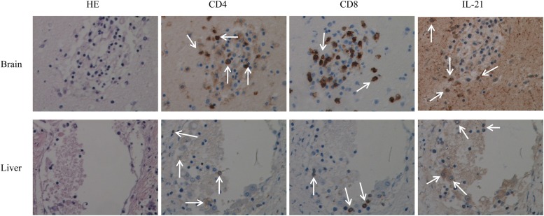
Fig 4.
Tissue sections from the brain (upper panels) and liver (lower panels) were used in immunochemistry studies to assess the presence of IL-21-producing T cells. CD4+ and CD8+ T lymphocytes were found to infiltrate brain and liver tissues. IL-21-producing T cell subtypes are shown (arrows point to stained cells for each marker). A representative immunohistochemistry (IHC) image (magnification, ×40) is shown. HE, hematoxylin and eosin.

