Fig 12.
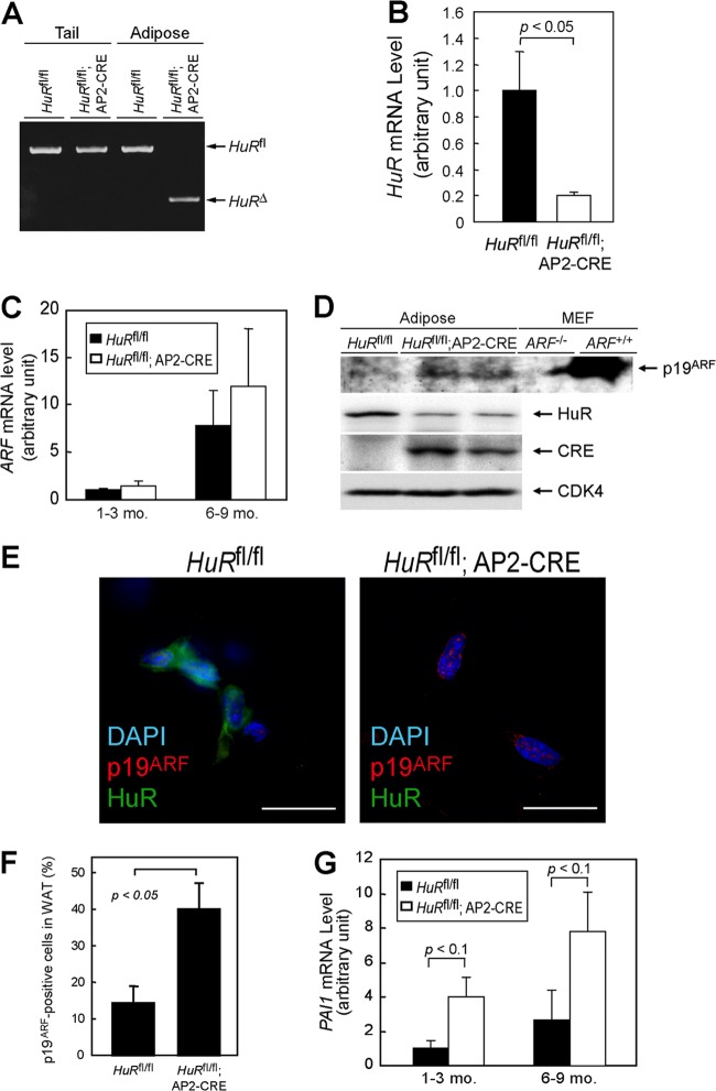
Adipose-specific HuR deletion accelerates the senescence of adipocytes. (A) Genotyping of adipose- and tail-derived genomic DNA. (B) HuR mRNA levels in adipose tissue from mice with the indicated genotypes were analyzed by real-time PCR. Values were normalized to 18S rRNA in each sample. (C) ARF mRNA was analyzed by real-time PCR. (D) Lysates were prepared from the adipose tissue of HuRfl/fl and HuRfl/fl; AP2-CRE mice. Expression of the indicated proteins was analyzed by immunoblotting. CDK4 was used as a loading control. Testis lysate from ARF−/− and ARF+/+ animals was used as the negative and positive controls for p19ARF, respectively. (E) Frozen sections of adipose tissue of HuRfl/fl and HuRfl/fl; AP2-CRE mice (9 months old) were immunostained using p19ARF and HuR antibodies. Sections were counterstained with DAPI. Bars, 20 μm. (F) Rates of p19ARF-positive cells in panel E were plotted. WAT, white adipose tissue. (G) PAI-1 mRNA levels were analyzed using real-time PCR.

