Fig 4.
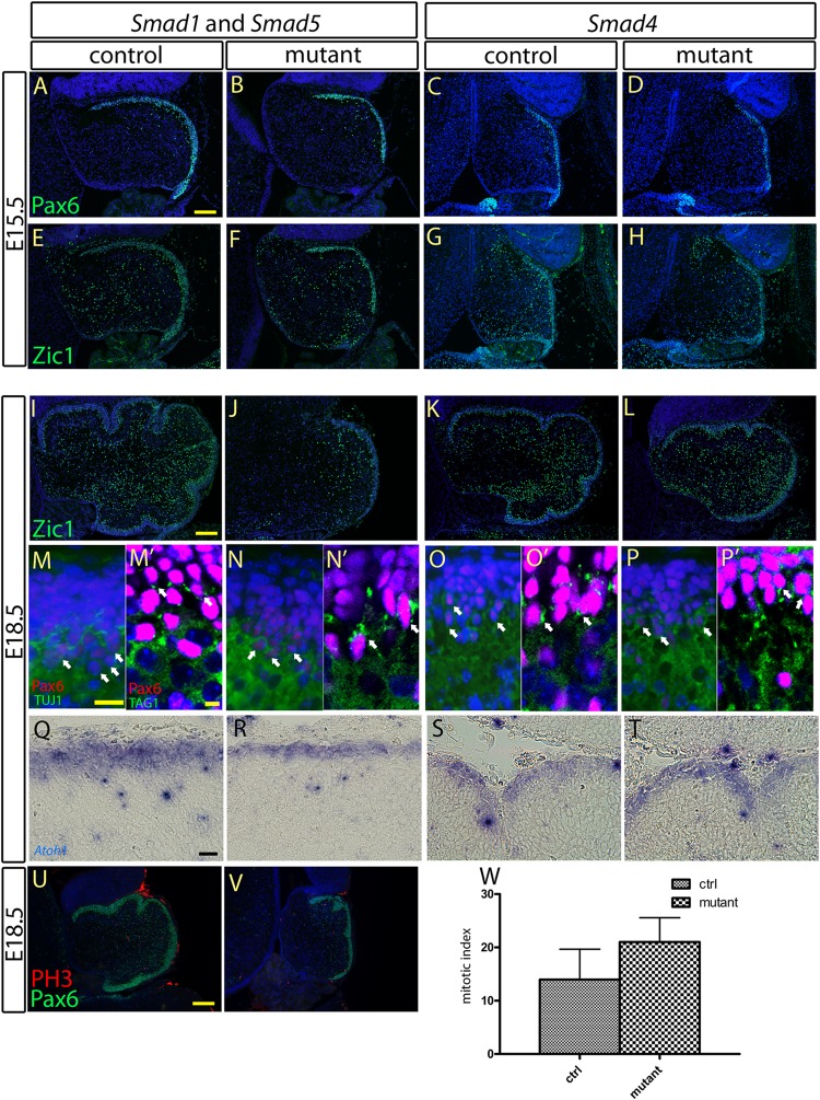
Characterization of the specification, differentiation, and maturation processes of the granule cell precursors at the EGL. (A to D, M to P, and Q to T) Immunostaining and in situ hybridization of the cerebellar sagittal sections at E15.5 and E18.5 showed normal expression of Pax6 and Atoh1, respectively, at the EGLs of the Smad1/5 mutant and the Smad4 mutant, indicating normal specification of the granule cell precursors at the EGL. (E to L) Immunostaining of Zic1 revealed no detectable abnormality in the differentiation process of granule cell precursors at the EGLs of the Smad1/5 mutant and the Smad4 mutant. Normal expression of Zic1 was detected in the cerebellar sagittal sections from both the controls and mutants at E15.5 (E to H) and E18.5 (I to L). (M to P′) Co-immunofluorescence staining of Pax6 with TUJ1 (M to P) and of Pax6 with TAG1 (M′ to P′) in the EGL sagittal sections of the controls, the Smad1/5 mutant, and the Smad4 mutant E18.5 cerebella showed coexpression of Pax6 with TUJ1 and TAG1 in granule cells, indicating normal maturation of granule cells from EGLs in both the Smad1/5 mutant and the Smad4 mutant. (U and V) Co-immunofluorescence staining of Pax6 and phospho-histone H3 (PH3) in sagittal sections of the control and Smad1/5 mutant E18.5 cerebella showed the cell-proliferating activity of the EGL. (W) No significant difference in cell proliferation of the EGL was detected between the control and Smad1/5 mutant at E18.5 (P = 0.0854). The error bars indicate standard deviations. Scale bars: 100 μm (A and I), 20 μm (M and Q), 5 μm (M′), and 150 μm (U).

