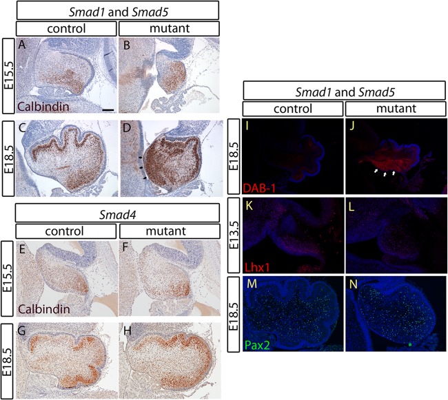
Fig 6.
A migration defect of Purkinje neurons in the cerebellum of the Smad1/5 mutant is related to reelin deficiency. (A to H) Immunostaining of calbindin in cerebellar sagittal sections at E15.5 and E18.5 from both the Smad1/5 mutant and the Smad4 mutant with their respective controls showed a migration defect in a population of Purkinje neurons of the Smad1/5 mutant that remains in the region near the VZ (D, arrows). Normal Purkinje cell migration was observed in the Smad4 mutant. (I and J) Immunostaining of DAB-1 in the E18.5 cerebellar sagittal sections showed overexpression of DAB-1 in the Purkinje cell population remaining near the VZ (arrows), indicating a defect in cerebellar reelin signaling of the Smad1/5 mutant. (K and L) Immunostaining of Lhx1 in E13.5 cerebellar sagittal sections showed a normal Lhx1 expression domain in the VZ of the Smad1/5 mutant, indicating a normal specification process of the VZ in the Smad1/5 mutant cerebellum. (M and N) Normal GABAergic neuron generation from the VZ of the Smad1/5 mutant shown by immunostaining of Pax2 in the E18.5 cerebellar sagittal sections. Scale bar, 100 μm.

