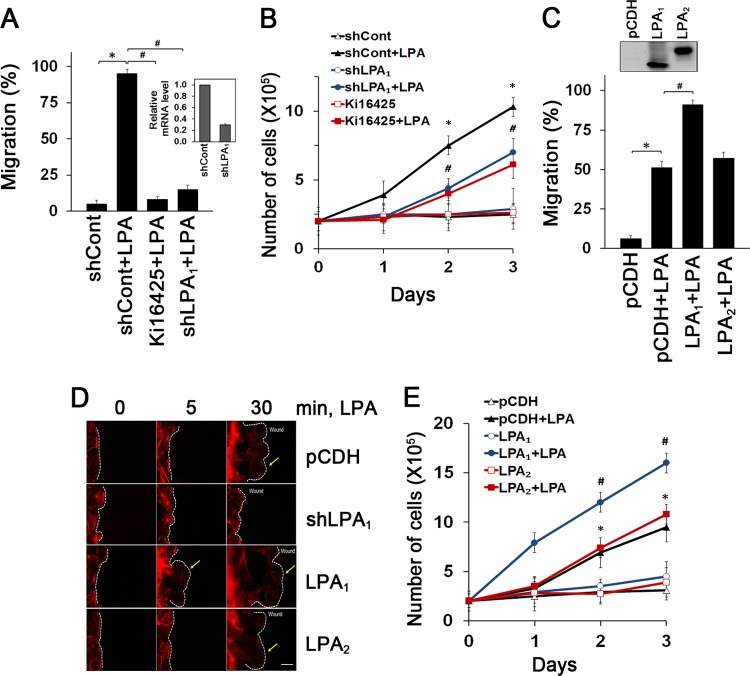
Fig 1.
Enhanced intestinal epithelial cell migration and proliferation are LPA1 dependent. (A) Migration of YAMC cells into the nude area at 24 h of 1 μM LPA treatment is shown. Cells were pretreated with 10 μM Ki16425 or transfected with shLPA1 prior to LPA treatment. The extent of wound closure was quantified. Full recovery of the wound was considered 100%. *, P < 0.001; #, P < 0.001. n = 3. BSA (0.1%) was used as a control. The inset shows LPA1 knockdown efficacy determined by real-time RT-PCR (shLPA1; 69% decrease). (B) LPA-induced cell proliferation was determined by counting cells for up to 3 days. Cells were pretreated with Ki16425 or transfected with shLPA1 as indicated. *, P < 0.01 versus shCont; #, P < 0.01 versus shCont plus LPA. Error bars represent the means ± SEM from three independent experiments involving triplicates. (C) The extent of migration at 12 h of LPA treatment was determined. *, P < 0.01; #, P < 0.01. n = 4. The inset shows overexpression of LPA1 and LPA2 in YAMC cells. (D) Lamellipodial extrusions in cells transfected with pCDH, shLPA1, LPA1, or LPA2 are shown. Cells were fixed and labeled with phalloidin-Alexa Fluor 568 to identify the leading edge of lamellipodia (arrows). Dashed lines indicate the leading edges. n = 3. Bar, 20 μm. (E) Numbers of proliferating cells transfected with LPA1 or LPA2 were determined. *, P < 0.01 versus pCDH; #, P < 0.01 compared with pCDH plus LPA. n = 3.

