Fig 2.
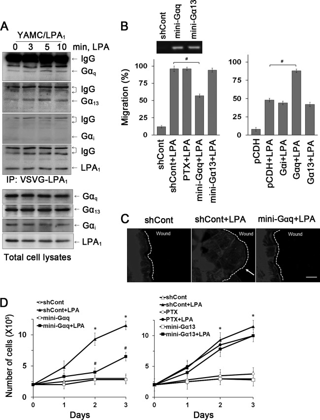
Gαq is essential for LPA1-mediated cell migration and proliferation. (A) The interaction of Gα subunits with LPA1 in YAMC/LPA1 was determined by coimmunoprecipitation of LPA1 and Gα subunits (top four panels). Expression of Gα subtypes in cell lysates is shown in the bottom panels. n = 3. (B) The role of Gα subtypes in cell migration was determined. YAMC cells pretreated with PTX or transfected with Gα minigenes were treated with LPA for 24 h (left panel). The inset shows the presence of Gα minigenes in transfected cells. The right panel shows the effect of Gα subunit overexpression on migration during 12 h of LPA treatment. #, P < 0.01. n = 3. Full recovery of the wound was considered 100%. #, P < 0.01. n = 3. (C) Lamellipodial extrusions labeled with phalloidin-Alexa Fluor 568 are shown. Dashed lines indicate the leading edges. n = 3. Bar, 20 μm. (D) Effects of inhibition of G proteins on proliferation are determined. (Left) Inhibition of Gαq. (Right) Inhibition of Gαi or Gα13. n = 3. *, P < 0.01 compared with shCont; #, P < 0.01 versus shCont plus LPA. n = 3.

