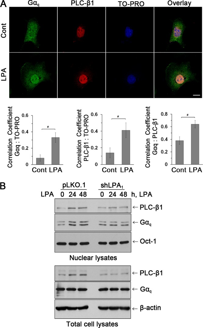
Fig 5.

LPA increased the nuclear abundance of Gαq and PLC-β1. (A) Expression of Gαq (green) and PLC-β1 (red) in the nuclei of YAMC cells was assessed by confocal immunofluorescence microscopy. TO-PRO iodide was used for nuclear counterstaining (blue). n = 3. Bars, 20 μm. Graphs represent Pearson's coefficient of colocalization of Gαq and TO-PRO (left), PLC-β1 and TO-PRO (middle), and Gαq and PLC-β1 (right) from 10 independent fields of cells. #, P < 0.05. (B) Western blot of Gαq and PLC-β1 in nuclear and total cell extracts. The transcription factor Oct-1 was used as a loading control for nuclear proteins. n = 3.
