Fig 2.
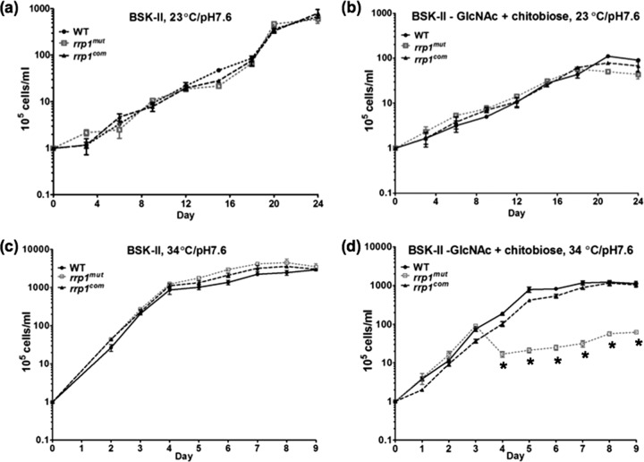
The growth curves of WT, rrp1mut, and rrp1com strains. Growth curves were measured under the following conditions: (a) cells were grown in BSK-II medium at 23°C and pH 7.6; (b) cells were grown in BSK-II(−GlcNAc)+chitobiose medium at 23°C and pH 7.6; (c) cells were grown in BSK-II medium at 34°C and pH 7.6; and (d) cells were grown in BSK-II(−GlcNAc)+chitobiose medium at 34°C and pH 7.6. Asterisks indicate that the difference in cell densities between the rrp1mut mutant and the WT was statistically significant at a P value of <0.001. Cell counting was repeated in triplicate with at least two independent samples, and the results are expressed as means ± SEM.

