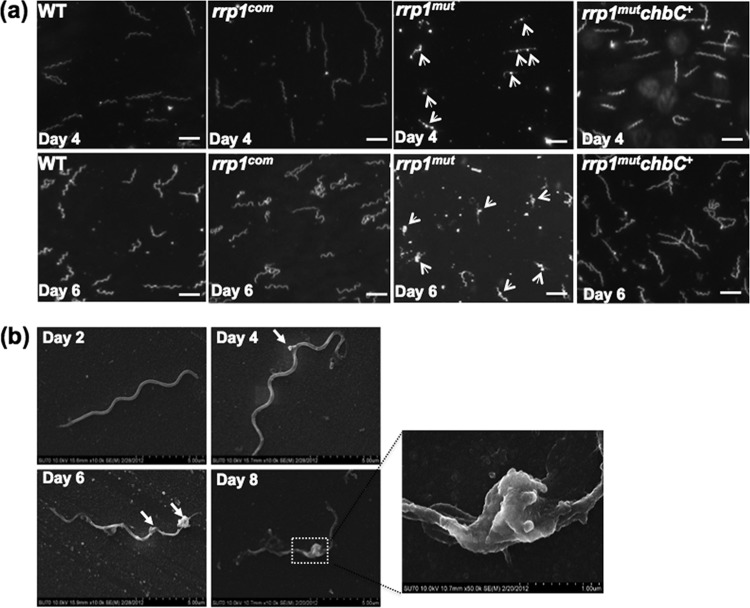
Fig 3.
The rrp1mut mutant forms membrane blebs in chitobiose-supplemented medium. (a) Dark-field microscopic analysis of B. burgdorferi strains grown in BSK-II(−GlcNAc)+chitobiose medium at 34°C and pH 7.6. The cultures were collected at day 4 and day 6 and B. burgdorferi cells observed under dark-field illumination at ×200 magnification using a Zeiss Axiostar Plus microscope. Scale bars represent 10 μm. (b) Scanning electron microscope analysis of the rrp1mut membrane bleb formation. A total of 105 cells/ml of the rrp1mut mutant were inoculated into 5 ml BSK-II(−GlcNAc)+chitobiose medium, and cells were collected for scanning electron microscope analysis at the different time points indicated to observe the formation of membrane blebs. Arrowheads point to membrane blebs observed.

