Fig 3.
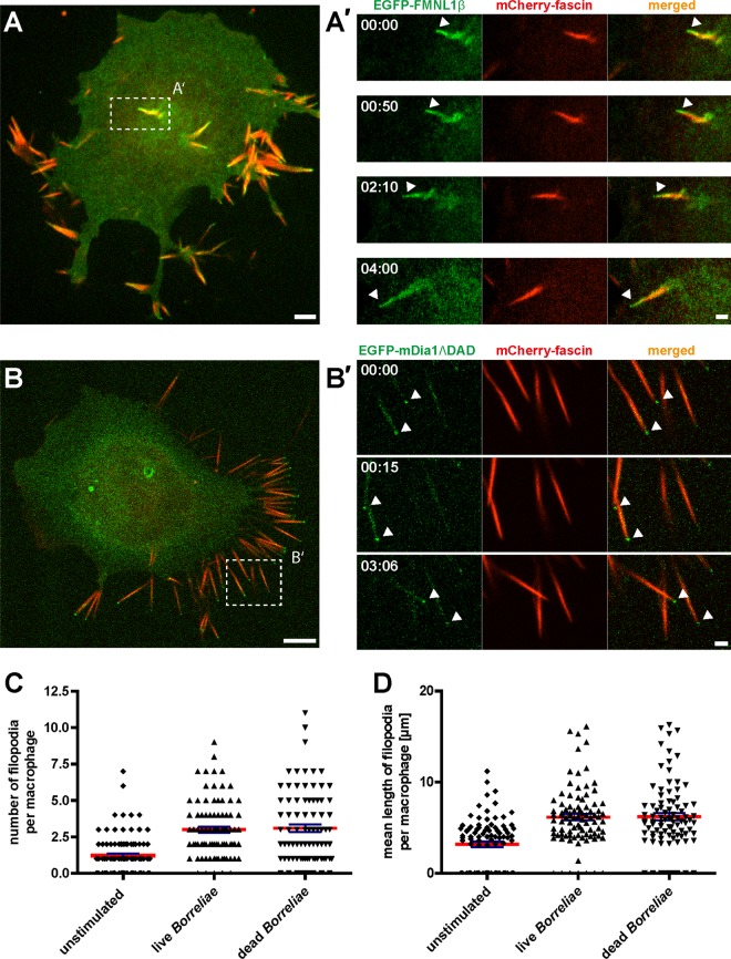
Borrelia-stimulated macrophage pseudopodia are enriched in EGFP-FMNL1β or EGFP-mDia1ΔDAD and are positive for the filopodium marker fascin. Confocal micrographs of macrophages expressing mCherry-fascin (red) and EGFP-FMNL1β (green; A) or EGFP-mDia1ΔDAD (green; B). (A and B) Still images from Videos S2 and S3 in the supplemental material. Dashed white boxes indicate the areas enlarged in the images shown in panels A′ and B′. Note localization of EGFP-FMNL1β along the length of filopodia, including the fascin-free tip (arrowhead), and its persistence over time (A′). Note the localization of EGFP-mDia1ΔDAD especially at the tips of filopodia (arrowheads) (B′). Scale bar, 2 μm. Time from the start of the experiments is indicated as min:sec. (C and D) Evaluation of number and length of filopodia in unstimulated macrophages and macrophages stimulated by addition of either live or heat-killed borreliae. To distinguish between microspikes and filopodia, only protrusions with a length of >3 μm were evaluated. Values are given as means ± SEM.

