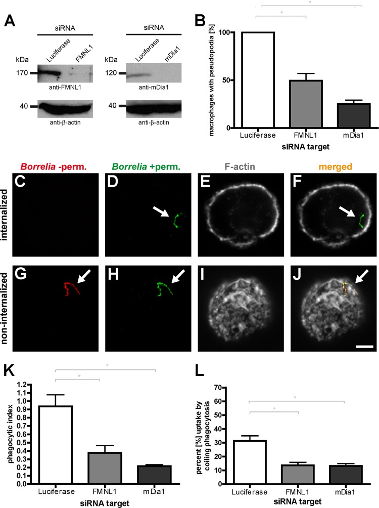
Fig 4.
siRNA-induced depletion of FMNL1 or mDia1 impairs the internalization of B. burgdorferi by primary human macrophages. (A) Western blots of lysates from macrophages treated with luciferase-specific siRNA (as a control), FMNL1-specific siRNA (left blot) or mDia1-specific siRNA (right blot). Proteins were detected with specific antibodies, with β-actin used as a loading control. Molecular mass in kDa is indicated. (B) Quantification of Borrelia-induced pseudopodia. The number of macrophages with pseudopodia in control cells treated with luciferase siRNA was set to 100%. Note the pronounced reduction of pseudopodium formation in cells depleted of FMNL1 or mDia1. Values are shown as means ± SEM (*, P < 0.05). The proportions of macrophages with pseudopodia are 49.6% ± 7.6% for FMNL1 siRNA and 25.1% ± 4.2% for mDia1 siRNA. For each value, each time 30 cells from three different donors were evaluated in three independent experiments. (C to J) Principle of outside-inside staining. Confocal laser scanning micrographs of macrophages coincubated with borreliae. To distinguish between internalized and noninternalized spirochetes, specimens were fixed and not permeabilized (−perm), stained with Bss42 B. burgdorferi antibody and with Alexa Fluor 568-labeled secondary antibody (red; C and G), and subsequently permeabilized (+perm) and stained with the same primary antibody but with Alexa Fluor 488-labeled secondary antibody (green; D and H). Internalized borreliae are detected only by the second round of staining and appear green; borreliae on the outside of the macrophages are detected by both stainings (red and green) and appear yellow in the merged image (F and J). Macrophages were stained with Alexa Fluor 647-labeled phalloidin to detect F-actin (E and I). Scale bar, 5 μm. (K) Internalization of borreliae by macrophages treated with specific siRNAs, based on evaluation of respective outside-inside staining. The phagocytic index is indicated as the ratio of internalized to noninternalized spirochetes. Values are given as means ± SEM (*, P < 0.05). Note the pronounced reduction of the phagocytic index upon depletion of either FMNL1 or mDia1 (phagocytic index of 0.38 ± 0.1 for FMNL1 siRNA and 0.22 ± 0.02 for mDia1 siRNA). For each value, each time 30 cells from three different donors were evaluated in three independent experiments. (L) Coiling phagocytosis of borreliae is reduced upon knockdown of FMNL1 or mDia1. Fixed specimens of borreliae in contact with macrophages were evaluated for the presence of several, clearly recognizable whorls enwrapping spirochetes as an indicator for coiling phagocytosis. Values are given as means ± SEM (*, P < 0.05). The percentages of uptake by coiling phagocytosis were 31.2% ± 6.5% for control macrophages transfected with luciferase siRNA, 13.7% ± 3.7% for macrophages transfected with FMNL1-specific siRNA, and 13.3% ± 2.9% for macrophages transfected with mDia1-specific siRNA. For the numbers of attached spirochetes per macrophage cell, see Fig. S3 in the supplemental material.

