
Fig 2.
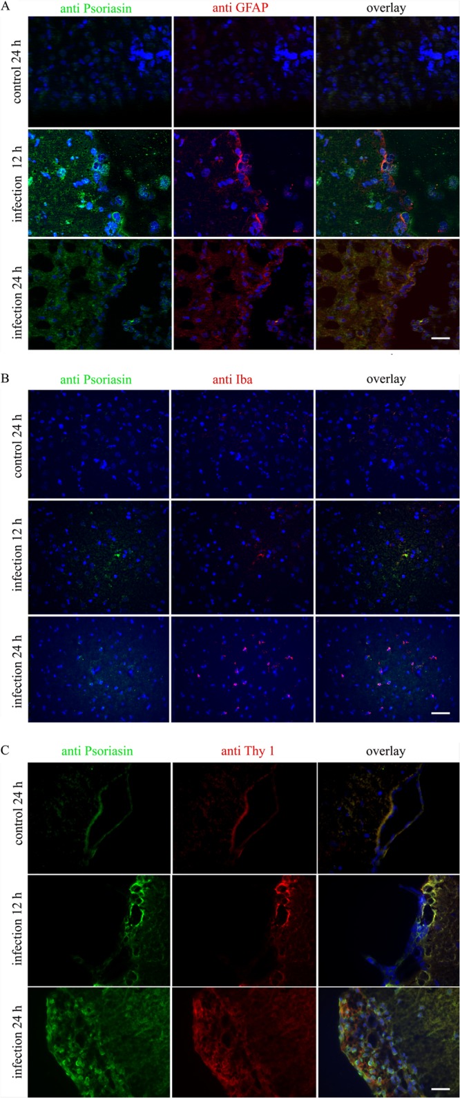
S100A15 expression and colocalization in an infant rat model of pneumococcal meningitis. Rat S100A15 expression in glial and meningeal cells was detected through immunohistochemistry in the infant rat model of pneumococcal meningitis. Juvenile rats were infected intracisternally with Streptococcus pneumoniae and sacrificed after 0, 12, or 24 h. The uninfected controls were injected with sterile saline. Coronal brain sections were fixed and immunolabeled with specific antibodies and examined with double-fluorescence microscopy. (A) Coronal brain sections were immunolabeled using anti-S100A15 (green) and anti-GFAP (red) to identify astrocytes and with DAPI for nuclear counterstaining (blue). (B) Coronal brain sections were immunolabeled using anti-S100A15 (green) and anti-Iba-1 (red) to identify microglia and with DAPI for nuclear counterstaining (blue). (C) Coronal brain sections were immunolabeled using anti-S100A15 (green) and anti-Thy-1 (red) to identify meningeal cells and with DAPI for nuclear counterstaining (blue). Coronal brain sections of three animals in triplicate were examined. Scale bar = 20 μm.