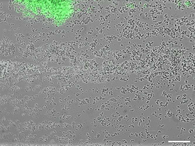
Fig 4.

High-level cid expression in small towers. S. aureus cells containing the cid::gfp reporter plasmid were inoculated into a BioFlux microfluidic system and allowed to form a biofilm as described in the legend to Fig. 3. The image shown (at ×200 magnification) represents a typical constitutive highly fluorescent “small” tower that is formed by this strain, distinct from the delayed fluorescence produced by the “large” towers depicted in Fig. 3. Note the presence of detached, highly fluorescent cells “downstream” (to the right) of the tower. The scale bar represents 50 μm.
