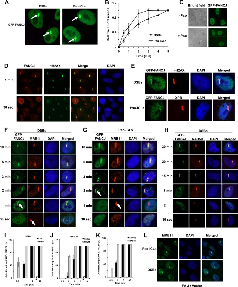
Fig 2.
Recruitment kinetics of FANCJ and MRE11 to laser-induced DSBs or psoralen cross-links. (A) GFP-FANCJ-transfected HeLa cells were treated with a laser (5.5%) at defined regions to induce localized DSBs or incubated with psoralen followed by laser treatment (1.8%) to induce Pso-ICLs. GFP-FANCJ recruitment to Pso-ICLs or DSBs was observed by live-cell imaging using confocal immunofluorescence microscopy. Arrows indicate FANCJ recruitment to laser-induced DSBs or Pso-ICLs, as specified. (B) Graphical representation of GFP-FANCJ recruitment to sites of DSBs or Pso-ICLs from 0 to 5 min after laser-induced damage, as detected by live-cell imaging. Relative fluorescence values expressed as a function of time are means for three independent experiments, with SD indicated by error bars. (C) Live-cell imaging of GFP-FANCJ recruitment to laser-irradiated GFP-FANCJ-transfected HeLa cells in either the presence or absence of psoralen. Black lines in the bright-field images indicate the defined areas irradiated with the laser. (D) Laser-targeted untransfected HeLa cells were stained with anti-FANCJ antibody and anti-γH2AX antibodies as described in Materials and Methods. (E) Laser-targeted GFP-FANCJ-transfected HeLa cells were stained with anti-γH2AX and anti-XPB antibodies as positive controls for DSBs and Pso-ICLs, respectively. (F and G) GFP-FANCJ-transfected HeLa cells were targeted with a laser at specific time points to induce DSBs (F) or Pso-ICLs (G), and cells were fixed and stained with anti-MRE11 antibody. (H) GFP-FANCJ-transfected HeLa cells were targeted with a laser at specific time points to induce DSBs, and cells were fixed and stained with anti-RAD50 antibody. (I and J) Graphical representation of GFP-FANCJ and MRE11 recruitment to sites of DSBs (I) or Pso-ICLs (J) from 0.5 to 10 min after laser-induced damage. (K) Graphical representation of GFP-FANCJ and RAD50 recruitment to sites of DSBs from 0.5 to 30 min after laser-induced damage. (L) FA-J cells were targeted with a laser to create either DSBs or Pso-ICLs and then were fixed 2 min after laser irradiation, followed by staining with anti-MRE11 antibody. For each experimental point, 20 to 25 cells were examined.

