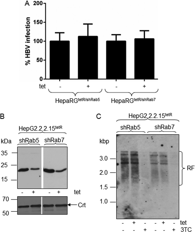
Fig 6.
HBV replication in Rab-depleted cells. Differentiated HepaRGtetR/shRab5 and HepaRGtetR/shRab7 cells were infected with 104 HBV Geq/cell and then treated with 1 μg/ml Tet from day 7 to day 14 p.i. (+) or maintained untreated (−) as the control. Infected cells were harvested 14 days p.i., and the amount of encapsidated viral DNA was quantified by real-time PCR. The results are expressed as percentage of HBV infection from untreated controls. The error bars represent the standard deviations among three independent experiments, each run in duplicate samples (A). HepaG2.2.2.15tetR/shRab5 and HepaG2.2.2.15tetR/shRab7 cells were induced with 1 μg/ml Tet for 3 days or maintained untreated as a control. Depletion of Rab5 and Rab7 was confirmed by Western blotting using specific Abs and Crt expression as a loading control (B). HepG2.2.2.15tetR/shRab5 and HepG2.2.2.15tetR/shRab7 cells were treated with 1 μg/ml Tet or 10 μM 3TC for 3 days or maintained untreated as a control. HBV nucleocapsids were extracted from an equal number of cells, and the purified DNA was analyzed by Southern blotting. The HBV replication forms (RF) were detected with a fluorescein-labeled probe obtained by random priming using the HBV DNA genome as the template (C).

