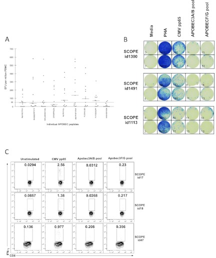
Fig 1.
APOBEC3-specific T cell responses in HIV-1-infected children. (A) PBMC from 13 chronically HIV-1-infected children were screened against 9 of the 12 APOBEC3 peptides present in the APOBEC3 peptide pool. Results are shown as the mean numbers of spot-forming units (SFU) per million PBMC of duplicate wells in the IFN-γ ELISPOT, and background SFUs have been subtracted from the values. The sequences of the individual peptide epitopes are shown on the x axis. (B) Examples of wells from an IFN-γ ELISPOT assay. One hundred thousand PBMC from three chronically infected adults were left unstimulated (Media) or stimulated with phytohemagglutinin (PHA), CMV pp65 peptide pool, APOBEC3A/B peptide pool, or APOBEC3F/G peptide pool. The number of spot-forming units/105 cells are indicated in the lower left corner for each well. (C) IFN-γ production by the CD8+ lymphocyte population from PBMC isolated from three chronically HIV-1-infected patients from the SCOPE cohort. The percentage of cells within the gate is shown in each plot: unstimulated, stimulated with a pool of CMV pp65 peptides, stimulated with a pool of APOBEC3A/B peptides, and stimulated with APOBEC3F/G pool.

