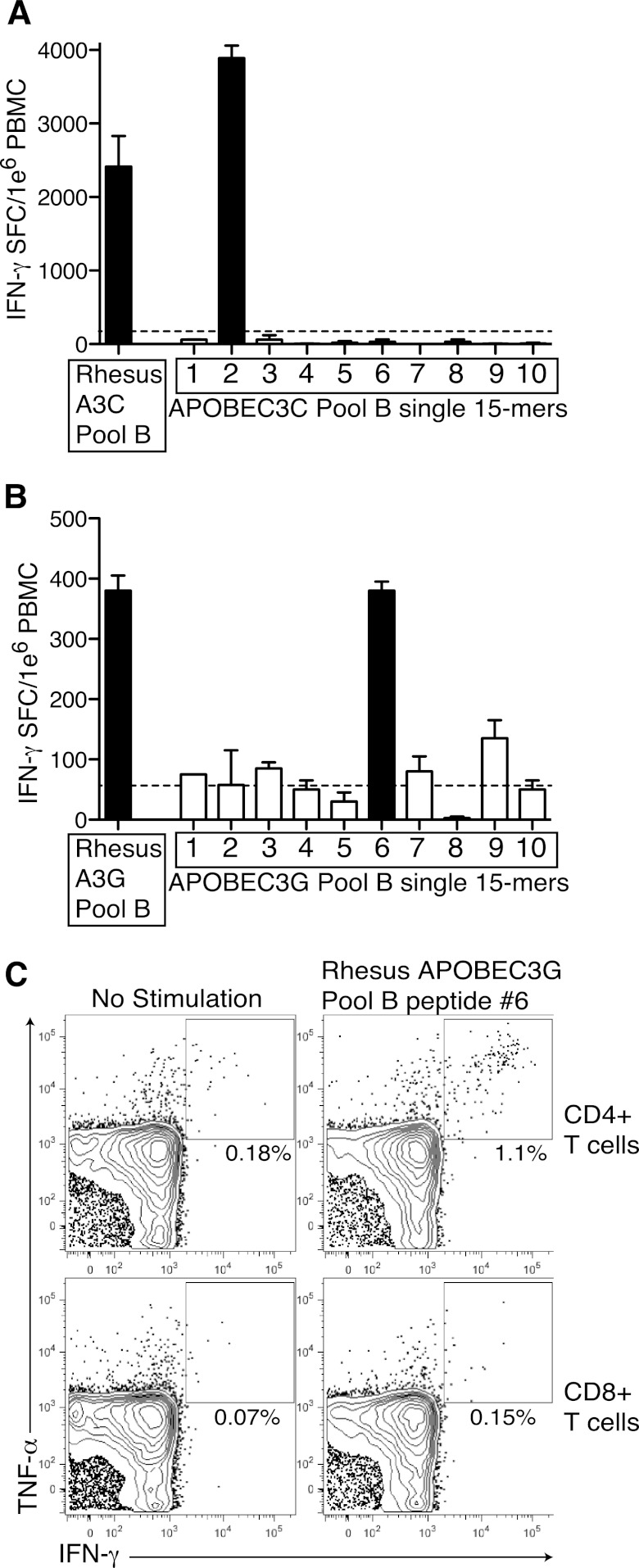
Fig 3.
Confirmation of APOBEC3-specific responses in SIV-infected rhesus macaques. (A) PBMC from LTNP rhesus macaques were screened against the APOBEC3C pool B and each individual 15-mer peptide within that pool. Results are shown as means plus standard deviations (error bars) of duplicate wells. (B) PBMC from LTNP rhesus macaques were screened against the APOBEC3G pool B and each individual 15-mer peptide within that pool. Black bars indicate positive responses. Results are shown as means plus standard deviations of duplicate wells. The broken line represents the threshold for positivity. (C) Bronchoalveolar lavage cells were stimulated with media (no stimulation) or the rhesus APOBEC3G pool B single 15-mer peptide identified in panel B. The percentages of cells positive for IFN-γ and tumor necrosis factor alpha (TNF-α) are shown.

