Fig 1.
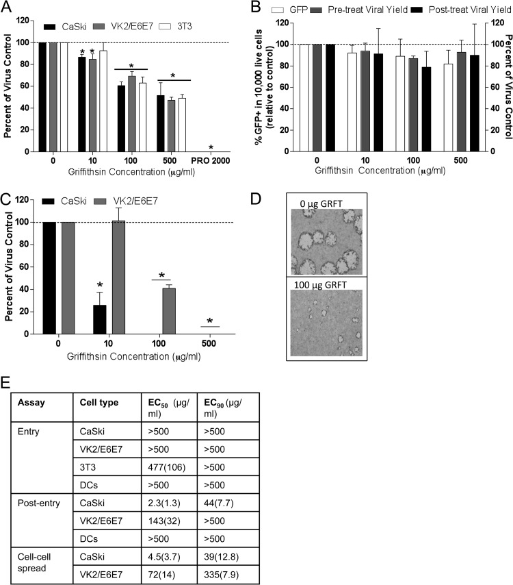
Griffithsin inhibits HSV-2 infection post entry. (A) CaSki, VK2/E6E7, and 3T3 cells were exposed to GRFT or PRO 2000 for 15 min, followed by challenge with HSV-2(4674) for 1 h. The cells were washed and then overlaid with medium, and plaques were counted after 24 to 48 h; results are presented as PFU/well as percentages of control (no drug) and are means + SEM from 2 to 4 experiments conducted in duplicate. (B) Immature moDCs were exposed to GRFT overnight and then challenged with HSV-2(333)ZAG (MOI of 1 PFU/cell) for 1 h at 37°C. Infection was assessed by quantifying GFP expression (10,000 live event acquisition) by flow cytometry 4 hpi. Results are presented as percentage of GFP+ cells and are means + SEM from six (pretreat) independent experiments. To monitor effects of GRFT on the release of viral progeny, the DCs were exposed to GRFT either before or after initial entry, and supernatants were harvested 24 hpi and overlaid onto Vero cells; viral plaques were counted after Giemsa staining and are presented as percentages relative to DCs infected in the absence of GRFT from 2 independent experiments, each conducted in duplicate. (C) CaSki or VK2/E6E7 cells were first exposed to HSV-2 for 1 h, washed with PBS, and then cultured in medium supplemented with the indicated concentration of GRFT; results are presented as percentages of the control and are the means + SEM from 3 experiments performed in duplicate. (D) Representative photograph of viral plaques in VK2/E6E7 cells when GRFT was added postentry. (E) EC50 and EC90 (μg/ml) were calculated for GRFT using data from panels A to C and Fig. 2A. Asterisks indicate P values of <0.05 compared to no drug (t test).

