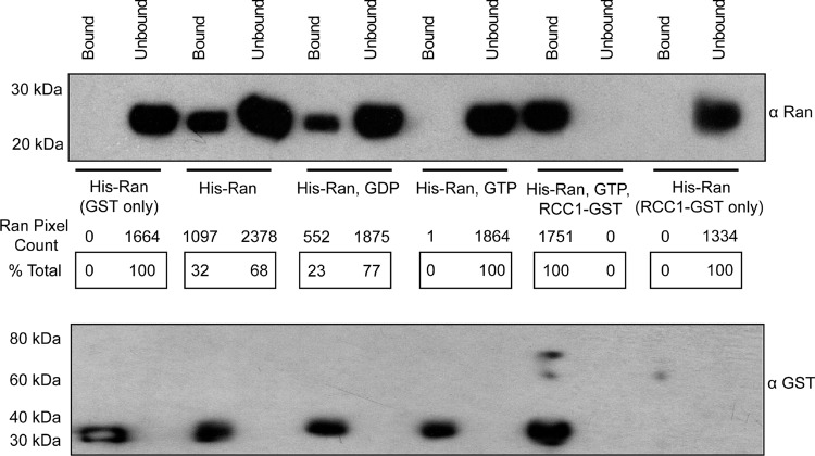
Fig 1.
GST-L–His-Ran binding. GST-L linked to glutathione-Sepharose beads (50 nM) was reacted (1 h, 25°C) with His-Ran (50 nM) in the presence or absence of GDP or GTP (2.5 μM) and/or RCC1-GST (1 nM). The clarified supernatant (Unbound) and bead-bound proteins (Bound) were precipitated (30% trichloroacetic acid), solubilized (alkaline SDS), fractionated by PAGE, and then transferred to polyvinylidene difluoride membranes. Western blot analyses used anti-Ran polyclonal antibody (product no. SC-1156; Santa Cruz Biotech) or anti-GST monoclonal antibody (product no. 71097; Novagen). Secondary antibodies were horseradish peroxidase-conjugated anti-goat antibody (product no. A5420; Sigma) or anti-mouse antibody (product no. A2554; Sigma). Relative pixel counts (% total) were determined by ImageQuant (GE Life Sciences) scanning of the membranes.

