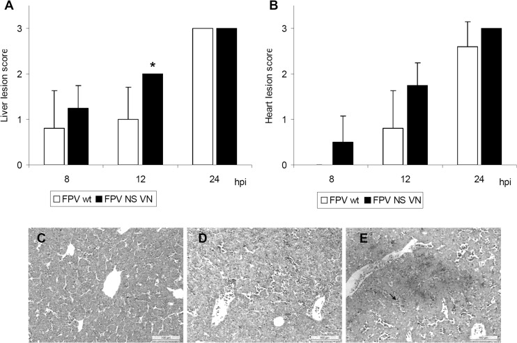
Fig 4.
Histopathologic examination of intravenously infected turkey embryos. Embryos were inoculated with 103 FFU of FPV wt or recombinant FPV NS VN HPAIV (n = 5/virus/time point). (A and B) Histopathologic lesions of liver and heart were evaluated using following lesion scores (not done for chicken embryos): 0, no lesions; 1, mild lesions with focal inflammation (edema and bleeding); 2, moderate lesions with focal to multifocal inflammation and scarce lymphocytic infiltration; 3, severe lesions with disseminated inflammation, tissue degeneration, and massive lymphocytic infiltration. Error bars indicate the standard deviations. Asterisks indicate statistically significant differences between FPV wt and FPV NS VN groups (P < 0.05; Wilcoxon rank sum test). No lesions were observed in virus-free embryos. (C to E) Histology of turkey embryonic liver at 12 hpi showing representative pictures of noninfected (C), FPV-wt infected (D), and FPV NS VN-infected (E) groups. The arrow indicates lymphocyte aggregation.

