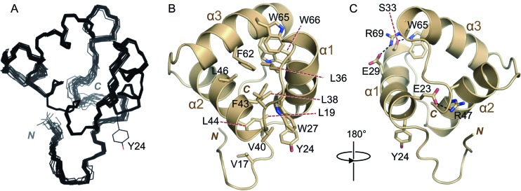
Fig 2.
Solution structure of FCV VPg 9-79. (A) Backbone trace of the 20 lowest-energy conformers of FCV VPg 9-79, calculated using ARIA (62) and CNS (60), including a final water refinement. The nucleotide-accepting tyrosine (Y24) side chain of one of the models is shown. (B) Representative conformer of FCV VPg 9-79, with selected (mainly hydrophobic) side chains shown as sticks and colored by atom type (tan, carbon; red, oxygen; blue, nitrogen). The N and C termini are indicated. (C) FCV VPg 9-79 rotated by 180° compared to panel B and with key charged and polar residues involved in electrostatic interactions shown as sticks. Polar interactions are indicated by dashed black lines.

