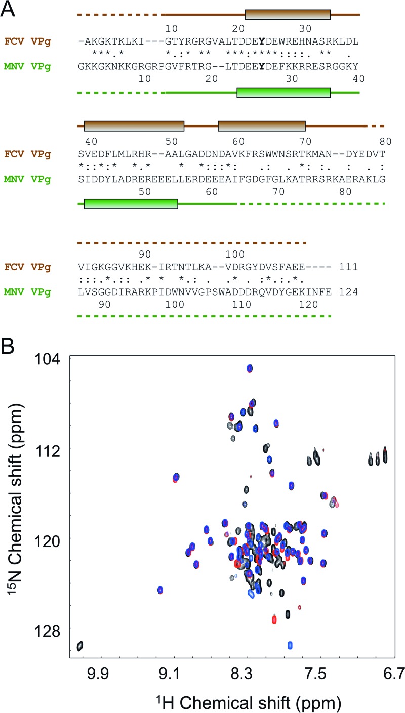
Fig 3.
NMR analysis of the MNV VPg benefitted from knowledge of the structure of FCV VPg. (A) Amino acid sequence alignment of FCV and MNV VPg proteins, performed using ClustalW. Positions of the helices detected through structural analysis are indicated as shaded boxes above and below the sequences (green for FCV and brown for MNV). Dashed lines indicate disordered portions of the polypeptide backbone. The nucleotidylated Tyr in each sequence is shown in boldface. Within the alignment, the asterisk, colon, and period characters denote identical, very similar, and similar amino acids, respectively. (B) Overlay of 1H-15N HQSC spectra of MNV VPg constructs used to probe the extent of the structure core of the protein: blue, VPg 11-85; black, VPg 1-124; red, VPg 11-62.

