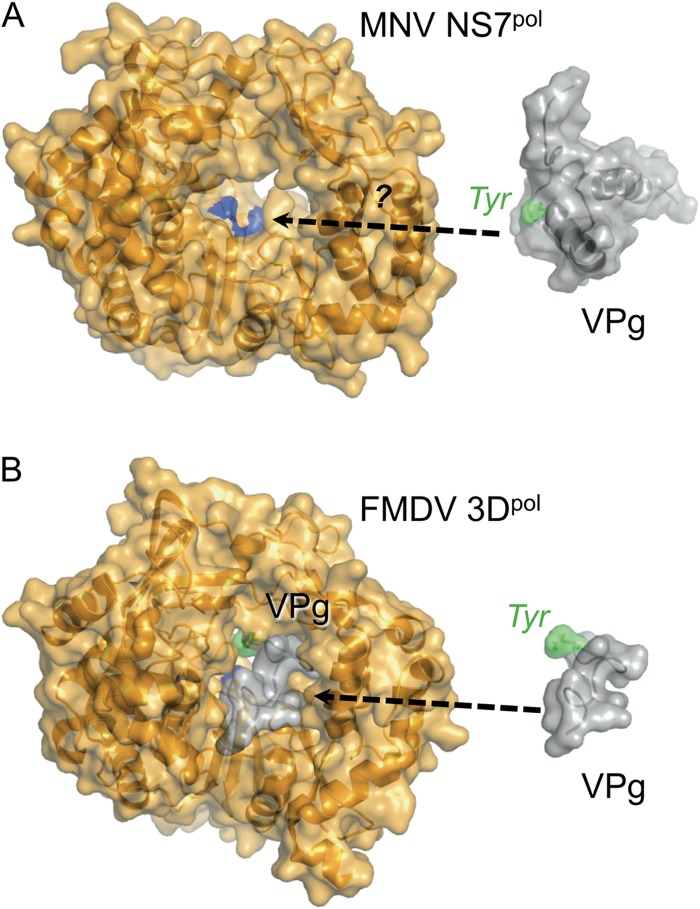
Fig 8.
How does MNV VPg interact with its polymerase? Comparison of the structures of polymerases and VPg proteins from MNV and FMDV. (A) Crystal structure of MNV NS7pol (67) and solution structure of MNV VPg 11-85 (this work). The active site of the polymerase, where nucleotidylation takes place, is colored blue. It is not yet clear what conformational changes are required for MNV VPg to be accommodated within the active site. (B) Cocrystal structure of FMDV 3Dpol and VPg (75), with VPg also shown extracted from the structure on the right (for ease of comparison with panel A). Only residues 1 to 15 of FMDV VPg were visible in the crystal structure.

