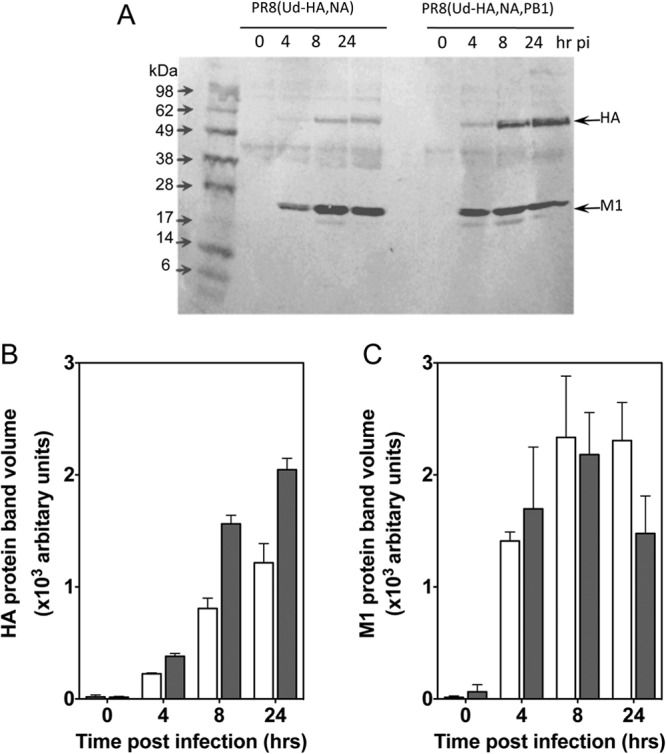
Fig 3.

Viral protein expression in cells infected with viruses differing in the origin of the PB1 protein. Aliquots of 1 × 10⁵ MDCK cells infected with PR8(Ud-HA,NA) virus or PR8(Ud-HA,NA,PB1) virus from the preparations used in the experiments whose results are shown in Figure 2B, C, and D were disrupted and separated on 4 to 20% Tris-glycine gels under nonreducing conditions. Proteins were transferred to PVDF membranes and probed with anti-Udorn HA and anti-M1 monoclonal antibodies. pi, postinfection. (A). The relative contents of HA (B) and M1 (C) proteins were determined by densitometry of stained bands and analyzed using the ImageQuant TL software. White bars, PR8(Ud-HA,NA) virus; gray bars, PR8(Ud-HA,NA,PB1) virus. Each bar represents the geometric mean of two independent experiments, and the error bars represent the standard errors of the means.
