
Fig 6.
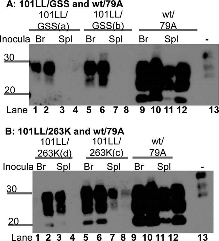
PrP-res levels in recipient mouse brains varied depending upon the tissue type inoculated from 101LL/GSS mice. This phenomenon was observed from both 101LL/GSS(a) and 101LL/GSS(b) mice (A). In contrast, 101LL/263K(c) and 101LL/263K(d) had varvarious levels of PrP-res independent of the tissue type inoculated (B), while wild-type/79A recipient brains had the same level of PrP-res independent of the tissue type inoculated (A and B). Tissue homogenates were PK digested and NaPTA precipitated. Lanes 1, 2, 5, 6, 9, and 10, brain tissue; lanes 3, 4, 7, 8, 11, and 12, spleen tissue. Samples were loaded at the equivalent of a wet tissue weight of 33 mg. An uninfected brain homogenate was loaded at 10 mg/ml (wt/vol) wet weight tissue in lane 13 as an internal control. Blots were probed with MAb 8H4. Numbers to the left of the panels indicate molecular weight markers (in kilodaltons).